
Procedure
Gasserian Ganglion Radiofrequency Ablation
Gasserian ganglion radiofrequency ablation targets the trigeminal nerve's sensory ganglion at the base of the skull to provide lasting relief from trigeminal neuralgia and severe facial pain.
Book a consultation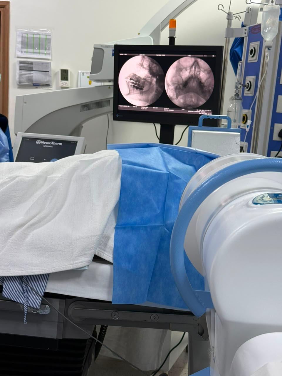
What is Gasserian Ganglion Radiofrequency Ablation?
The Gasserian ganglion (also called the trigeminal ganglion) is the sensory ganglion of the trigeminal nerve, located deep at the base of the skull in a recess called Meckel’s cave. It is the central relay point for all three divisions of the trigeminal nerve: the ophthalmic (V1), maxillary (V2), and mandibular (V3) branches, which together supply sensation to the entire face, scalp, sinuses, and upper teeth. When the trigeminal nerve is the source of severe, treatment-resistant facial pain, targeting the ganglion directly allows us to selectively disrupt pain-transmitting fibres at their origin.
The procedure is performed percutaneously (through the skin, with no incision) under fluoroscopic guidance. A fine needle electrode is advanced through a small opening at the base of the skull called the foramen ovale to reach the ganglion. Sensory stimulation at low frequency is then used to confirm the needle tip is adjacent to the division responsible for the pain: the patient reports where they feel the tingling. Once position is confirmed, a small radiofrequency lesion interrupts pain transmission in that division while sparing, as far as possible, the touch-sensing fibres that run alongside.
We offer both conventional radiofrequency — which uses heat at 65 to 80°C to create a durable lesion — and pulsed radiofrequency, which operates at 42°C in short bursts and modulates the nerve without destroying it. Pulsed RF is preferred when preserving more facial sensation is a priority. The choice depends on the severity of neuralgia, prior treatments, and the patient’s anatomy.
What conditions does it treat?
This procedure is primarily used for trigeminal neuralgia: the sharp, electric-shock pain in the face triggered by light touch, chewing, or speaking. It is offered when medication (carbamazepine, oxcarbazepine, pregabalin) has provided insufficient relief or caused intolerable side effects, and when the patient prefers or is better suited to a minimally invasive approach over open surgery. It is also effective for atypical facial pain with a clearly defined trigeminal distribution, and for post-herpetic neuralgia affecting the face after ophthalmic shingles.
What to expect
Before: A thorough facial pain assessment confirms the diagnosis and identifies which trigeminal division is involved. You will typically have tried at least one medication before this procedure. Blood thinners are paused in the days before. Light fasting (four to six hours) is required as the procedure is performed under short-acting sedation.
During: You lie on the procedure table with fluoroscopy guidance overhead. Local anaesthetic numbs the skin at the entry point on the cheek. Under live X-ray guidance, the needle is advanced through the foramen ovale to the ganglion — a precise, image-guided step that takes around 10 to 15 minutes. You are briefly brought out of sedation for stimulation testing, confirming the electrode is at the correct division before ablation. Each lesion takes 60 to 90 seconds. The full procedure typically takes 30 to 60 minutes.
After: You are observed for one to two hours and discharged the same day. Some facial soreness and numbness in the treated division are expected in the days following. Most patients experience significant pain relief within the first week. Relief typically lasts two to four years. If the neuralgia returns, the procedure can be repeated. A small proportion of patients develop lasting numbness or dysaesthesia — a troublesome sensation of painful numbness — and this risk is discussed in detail before you consent.
Conditions this treats
Related services









Tell us what's hurting.
One consultation. One coordinated team. A plan built around your case.